Ventricles of brain pdf Canterbury
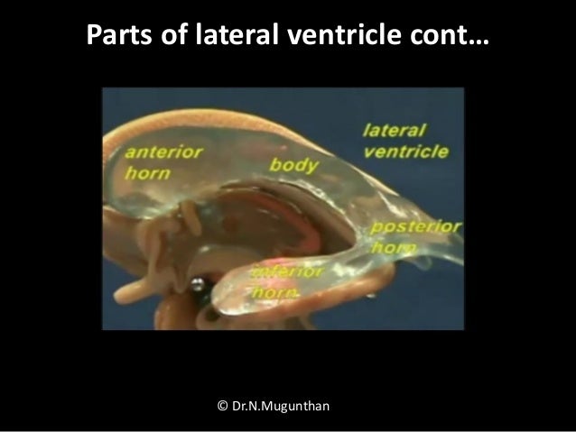
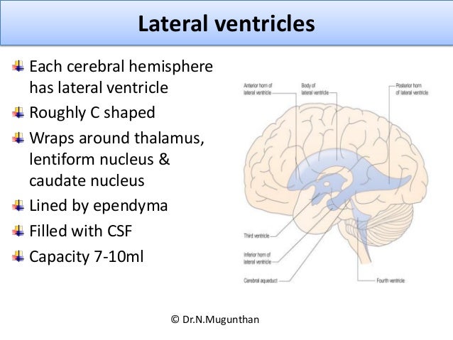
(PDF) The ventricular system of the brain A comprehensive The lateral ventricles are the two largest cavities of the ventricular system of the human brain and contain cerebrospinal fluid (CSF). Each cerebral hemisphere contains a lateral ventricle, known as the left or right ventricle, respectively.. Each lateral ventricle resembles a C-shaped structure that begins at an inferior horn in the temporal lobe, travels through a body in the parietal lobe
N6 – Ventricles of Brain
[PDF] The Ventricular System of the Brain Semantic Scholar. Brain ventricle: One of the communicating cavities within the brain. There are four ventricles: two lateral ventricles, the third ventricle, and the fourth ventricle. The lateral ventricles are in the cerebral hemispheres. Each lateral ventricle consists of a triangular central body and four horns, VENTRICLES OF THE BRAIN AND CSF - Free download as Powerpoint Presentation (.ppt), PDF File (.pdf), Text File (.txt) or view presentation slides online. A brief seminar given by myself to the class on the ventricles of the brain and CSF.
Hetzel W. The posterior horn and collateral trigone of the lateral ventricle of the monkey brain (Macaca speziosa). A scanning electron microscopic study. Cell Tissue Res. 1978 Jan 9; 186 (1):161–170. Hirunagi K, Yasuda M. Scanning electron microscopic analysis of the linings of the fourth ventricle in the domestic fowl. BI 335 – Advanced Human Anatomy and Physiology Western Oregon University Figure 4: Mid-sagittal section of brain showing diencephalon (includes corpus callosum, fornix, and anterior commissure) Marieb & Hoehn (Human Anatomy and Physiology, 9th ed.) – Figure 12.10 Exercise 2: Utilize the model of the human brain to locate the following structures / landmarks for the
Neurology: Ventricular system of the Brain. STUDY. PLAY. Human Brains-Brain is suspended in CSF and partially floats in the skull cavity -There are 4 cavities in the brain called ventricles. All 4 ventricles are interconnected through narrow-pipe like openings. CSF-is produced predominately in the brain ventricles -functions as a buffer to absorb stress transferred through the rigid skull 22/02/2015 · Ventricles of the brain: want to learn more about it? Our engaging videos, interactive quizzes, in-depth articles and HD atlas are here to get you top results faster. Sign up for your free Kenhub account today and join over 1,195,034 successful anatomy students.
22/02/2015 · Ventricles of the brain: want to learn more about it? Our engaging videos, interactive quizzes, in-depth articles and HD atlas are here to get you top results faster. Sign up for your free Kenhub account today and join over 1,195,034 successful anatomy students. cerebral aqueduct) into the fourth ventricle which lies towards the base of the brain. From the fourth ventricle, it flows around the spinal cord and over the surface of the brain before being re-absorbed. Without signs of increased pressure in the brain (hydrocephalus), ventriculomegaly most likely will not cause any problems. However, it can
VENTRICLES OF THE BRAIN AND CSF - Free download as Powerpoint Presentation (.ppt), PDF File (.pdf), Text File (.txt) or view presentation slides online. A brief seminar given by myself to the class on the ventricles of the brain and CSF cerebral aqueduct) into the fourth ventricle which lies towards the base of the brain. From the fourth ventricle, it flows around the spinal cord and over the surface of the brain before being re-absorbed. Without signs of increased pressure in the brain (hydrocephalus), ventriculomegaly most likely will not cause any problems. However, it can
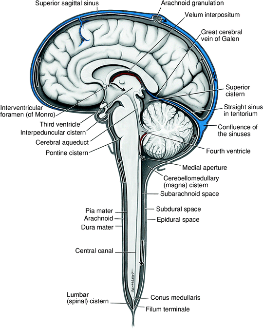
2.2.7 Ventricles. The ventricles of the brain circulate cerebrospinal fluid. There are two lateral ventricles and a third and fourth ventricle. These will each be described from a superior to inferior direction. The lateral ventricles (left and right) communicate with the third ventricle below via an interventricular foramen on each side. The ventricles and central canal of the spinal cord initially form a closed system. However, by about the third month of development, three openings form in the roof of the fourth ventricle, rendering the ventricular system continuous with the subarachnoid space surrounding the brain and spinal cord.
The ventricular system is the extension of the spinal canal (canalis centralis) into the brain and consists of four chambers which are filled with cerebrospinal fluid (liquor cerebrospinalis). The paired lateral ventricles (ventriculi laterales I and II) are two of these four chambers and are connected to the unpaired third and fourth ventricle through the foramen interventriculare. 2.2.7 Ventricles. The ventricles of the brain circulate cerebrospinal fluid. There are two lateral ventricles and a third and fourth ventricle. These will each be described from a superior to inferior direction. The lateral ventricles (left and right) communicate with the third ventricle below via an interventricular foramen on each side.
Ventricular system of brain Dr. Syed Imad FCPS, MRCS Start studying Ventricular System of the Brain. Learn vocabulary, terms, and more with flashcards, games, and other study tools.
30/06/2016 · The ventricles of the brain are a communicating network of cavities filled with cerebrospinal fluid (CSF) and located within the brain parenchyma. The ventricular system is composed of 2 lateral ventricles, the third ventricle, the cerebral aqueduct, and the fourth ventricle (see the following images). 30/06/2016 · The ventricles of the brain are a communicating network of cavities filled with cerebrospinal fluid (CSF) and located within the brain parenchyma. The ventricular system is composed of 2 lateral ventricles, the third ventricle, the cerebral aqueduct, and the fourth ventricle (see the following images).
The ventricles and central canal of the spinal cord initially form a closed system. However, by about the third month of development, three openings form in the roof of the fourth ventricle, rendering the ventricular system continuous with the subarachnoid space surrounding the brain and spinal cord. The Ventricular System of the Brain - Free download as Powerpoint Presentation (.ppt / .pptx), PDF File (.pdf), Text File (.txt) or view presentation slides online. cns
Slide 10 of 25 of Fourth ventricle The ventricular system is a series of connecting hollow spaces called ventricles in the brain that are filled with cerebrospinal fluid. The ventricular system consists of two lateral ventricles, the third ventricle, and the fourth ventricle. The cerebral ventricles are connected by small pores called foramina, as well as by larger channels.The interventricular foramina or foramina of …
The Ventricular System and Cerebrospinal Fluid (CSF)
The Ventricular System of the Brain Cerebrospinal Fluid. The Ventricular System and Cerebrospinal Fluid (CSF) Simple Tube Shape of Early CNS with fluid filled canal in middle In adults there is still a continuous canal running thru all levels of the adult CNS – it is just harder to follow. Book Fig. 1.19 Side & Frontal Views of Ventricles Remember – these represent fluid filled cavities in brain, 2.2.7 Ventricles. The ventricles of the brain circulate cerebrospinal fluid. There are two lateral ventricles and a third and fourth ventricle. These will each be described from a superior to inferior direction. The lateral ventricles (left and right) communicate with the third ventricle below via an interventricular foramen on each side..
VENTRICLES OF THE BRAIN AND CSF Cerebrospinal Fluid. The ventricular system is the extension of the spinal canal (canalis centralis) into the brain and consists of four chambers which are filled with cerebrospinal fluid (liquor cerebrospinalis). The paired lateral ventricles (ventriculi laterales I and II) are two of these four chambers and are connected to the unpaired third and fourth ventricle through the foramen interventriculare., REVIEW PAPER The ventricular system of the brain: a comprehensive review of its history, anatomy, histology, embryology, and surgical considerations.
On Serous Effusion from the Membranes and into the
Morphometric Analysis of the Brain Ventricles in Normal. Ventricular system of brain Dr. Syed Imad FCPS, MRCS https://simple.wikipedia.org/wiki/Category:Brain 2.2.7 Ventricles. The ventricles of the brain circulate cerebrospinal fluid. There are two lateral ventricles and a third and fourth ventricle. These will each be described from a superior to inferior direction. The lateral ventricles (left and right) communicate with the third ventricle below via an interventricular foramen on each side..

Brain –Introduction Figure 2 Normal islets of Calleja in a male B6C3F1 mouse from a 2-year study. The arrows identify the typical location and morphology of the islets of Calleja. Figure 2 shows the Islets of Calleja in the frontoparietal cortex.These are a common, normal occurrence of symmetrical clusters of primitive neural cell components in the ventral VENTRICLES OF THE BRAIN AND CSF - Free download as Powerpoint Presentation (.ppt), PDF File (.pdf), Text File (.txt) or view presentation slides online. A brief seminar given by myself to the class on the ventricles of the brain and CSF
Brain ventricle: One of the communicating cavities within the brain. There are four ventricles: two lateral ventricles, the third ventricle, and the fourth ventricle. The lateral ventricles are in the cerebral hemispheres. Each lateral ventricle consists of a triangular central body and four horns Ventricular system of brain Dr. Syed Imad FCPS, MRCS
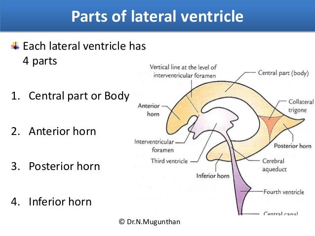
The Ventricular System and Cerebrospinal Fluid (CSF) Simple Tube Shape of Early CNS with fluid filled canal in middle In adults there is still a continuous canal running thru all levels of the adult CNS – it is just harder to follow. Book Fig. 1.19 Side & Frontal Views of Ventricles Remember – these represent fluid filled cavities in brain N7 Ventricles of Brain 1. left lateral ventricle 2. right lateral ventricle 3. anterior horn 4. inferior horn 5. posterior horn 6. median portion of lateral ventricle 7. communication between lateral ventricle and third ventricle 8. third ventricle 9. interthalamic adhaesion 10. portion of third ventricle reaching into pineal gland
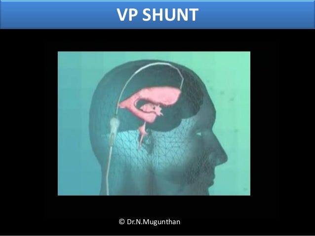
the brain. Each ventricle lies in contact with five critical neural structures: the caudate nucleus, the thalamus, the fornix, the corpus callosum, and the genu of internal capsule. The authors report their experience in primary tumors of the lateral ventricles of the brain … The most severe form of slit ventricle syndrome occurs in children. The absence of cerebrospinal fluid (CSF) within the ventricles combined with a growing brain leads to situation in which "the brain is too big for the skull." The intracranial pressure (brain pressure) can be very high.
Dandy W. E. Benign encapsulated tumors in the lateral ventricles of the brain. Diagnosis and treatment. Baltimore: Williams & Wilkins Co. 1934 viii 189 pp. Dandy W. E. Benign encapsulated tumors in the lateral ventricles of the brain. Diagnosis and treatment. VENTRICLES OF THE BRAIN AND CSF - Free download as Powerpoint Presentation (.ppt), PDF File (.pdf), Text File (.txt) or view presentation slides online. A brief seminar given by myself to the class on the ventricles of the brain and CSF
The ventricles of the heart function to pump blood to the entire body. During the diastole phase of the cardiac cycle, the atria and ventricles are relaxed and the heart fills with blood. During the systole phase, the ventricles contract pumping blood to the major arteries (pulmonary and aorta).The heart valves open and close to direct the flow of blood between the heart chambers … N7 Ventricles of Brain 1. left lateral ventricle 2. right lateral ventricle 3. anterior horn 4. inferior horn 5. posterior horn 6. median portion of lateral ventricle 7. communication between lateral ventricle and third ventricle 8. third ventricle 9. interthalamic adhaesion 10. portion of third ventricle reaching into pineal gland
The most severe form of slit ventricle syndrome occurs in children. The absence of cerebrospinal fluid (CSF) within the ventricles combined with a growing brain leads to situation in which "the brain is too big for the skull." The intracranial pressure (brain pressure) can be very high. The most severe form of slit ventricle syndrome occurs in children. The absence of cerebrospinal fluid (CSF) within the ventricles combined with a growing brain leads to situation in which "the brain is too big for the skull." The intracranial pressure (brain pressure) can be very high.
Slide 10 of 25 of Fourth ventricle the brain. Each ventricle lies in contact with five critical neural structures: the caudate nucleus, the thalamus, the fornix, the corpus callosum, and the genu of internal capsule. The authors report their experience in primary tumors of the lateral ventricles of the brain …
VENTRICLES OF THE BRAIN AND CSF - Free download as Powerpoint Presentation (.ppt), PDF File (.pdf), Text File (.txt) or view presentation slides online. A brief seminar given by myself to the class on the ventricles of the brain and CSF Neurology: Ventricular system of the Brain. STUDY. PLAY. Human Brains-Brain is suspended in CSF and partially floats in the skull cavity -There are 4 cavities in the brain called ventricles. All 4 ventricles are interconnected through narrow-pipe like openings. CSF-is produced predominately in the brain ventricles -functions as a buffer to absorb stress transferred through the rigid skull
Brain ventricle: One of the communicating cavities within the brain. There are four ventricles: two lateral ventricles, the third ventricle, and the fourth ventricle. The lateral ventricles are in the cerebral hemispheres. Each lateral ventricle consists of a triangular central body and four horns The present investigation examined the ependymal linings of the cerebral ventricles of the dog following a single intracisternal injection of the viable …
The ventricles are filled with cerebrospinal fluid (CSF) which bathes and cushions the brain and spinal cord within their bony confines. CSF is produced by modified ependymal cells of the choroid plexus found in all components of the ventricular system except for the cerebral aqueduct and the posterior and anterior horns of the lateral ventricles. Ventricular system, meninges and blood vessels of the . brain . Chapters 7 and 9 (Head and cranial nerves) and lecture/seminar material . GENERAL OBJECTIVES:-Understand the organization of protective layers of the brain. -Dural venous sinuses, infoldings/reflections and layers of dura mater. -Arachnoid and pia mater end meningeal spaces. -
VENTRICLES OF THE BRAIN AND CSF Cerebrospinal Fluid
Tumors of the Lateral Ventricles of the Brain in Journal. The Ventricles and Cerebrospinal Fluid. There are four cavities in the brain, called ventricles. The ventricles are filled with cerebrospinal fluid (CSF), which provides the following functions: Absorbs physical shocks to the brain. Distributes nutritive materials to and removes wastes from nervous tissue. Provides a chemically stable environment. There are four ventricles: Each of …, Hetzel W. The posterior horn and collateral trigone of the lateral ventricle of the monkey brain (Macaca speziosa). A scanning electron microscopic study. Cell Tissue Res. 1978 Jan 9; 186 (1):161–170. Hirunagi K, Yasuda M. Scanning electron microscopic analysis of the linings of the fourth ventricle in the domestic fowl..
The cerebral ventricles of the dog SpringerLink
Ventricles of the Brain YouTube. 16/11/2013 · Methods. The literature was searched for articles and textbooks of different topics related to the history, anatomy, physiology, histology, embryology and surgical considerations of the brain ventricles., The ventricles of the heart function to pump blood to the entire body. During the diastole phase of the cardiac cycle, the atria and ventricles are relaxed and the heart fills with blood. During the systole phase, the ventricles contract pumping blood to the major arteries (pulmonary and aorta).The heart valves open and close to direct the flow of blood between the heart chambers ….
N7 Ventricles of Brain 1. left lateral ventricle 2. right lateral ventricle 3. anterior horn 4. inferior horn 5. posterior horn 6. median portion of lateral ventricle 7. communication between lateral ventricle and third ventricle 8. third ventricle 9. interthalamic adhaesion 10. portion of third ventricle reaching into pineal gland 22/06/2013 · This brief animation illustrates the location of the ventricles within the brain and the associated structures around it. The structures were sculpted digitally using ZBrush and were combined with
VENTRICLES OF THE BRAIN AND CSF - Free download as Powerpoint Presentation (.ppt), PDF File (.pdf), Text File (.txt) or view presentation slides online. A brief seminar given by myself to the class on the ventricles of the brain and CSF The ventricles are filled with cerebrospinal fluid (CSF) which bathes and cushions the brain and spinal cord within their bony confines. CSF is produced by modified ependymal cells of the choroid plexus found in all components of the ventricular system except for the cerebral aqueduct and the posterior and anterior horns of the lateral ventricles.
BI 335 – Advanced Human Anatomy and Physiology Western Oregon University Figure 4: Mid-sagittal section of brain showing diencephalon (includes corpus callosum, fornix, and anterior commissure) Marieb & Hoehn (Human Anatomy and Physiology, 9th ed.) – Figure 12.10 Exercise 2: Utilize the model of the human brain to locate the following structures / landmarks for the 29/05/2015 · Ventricles of the Brain-+ Dailymotion. For You Explore. Do you want to remove all your recent searches? All recent searches will be deleted. Cancel Remove. Log in. Watch fullscreen. Ventricles of the Brain. Windowbluff
Start studying Ventricular System of the Brain. Learn vocabulary, terms, and more with flashcards, games, and other study tools. PDF Introduction: As the human brain ages, characteristic structural changes occur that are considered to be normal and are expected. Thus the thorough knowledge of …
The ventricular system is the extension of the spinal canal (canalis centralis) into the brain and consists of four chambers which are filled with cerebrospinal fluid (liquor cerebrospinalis). The paired lateral ventricles (ventriculi laterales I and II) are two of these four chambers and are connected to the unpaired third and fourth ventricle through the foramen interventriculare. 08/07/2016 · The interstitial spaces of the brain are filled with cerebrospinal fluid (CSF). Faubel et al. studied fluid transport in the third ventricle of the brain of mice, rats, and pigs. Sophisticated, state-of-the-art fluid dynamic studies revealed a complex pattern of cilia beating that leads to an intricate network of “highways” of CSF flow.
PDF Introduction: As the human brain ages, characteristic structural changes occur that are considered to be normal and are expected. Thus the thorough knowledge of … Hetzel W. The posterior horn and collateral trigone of the lateral ventricle of the monkey brain (Macaca speziosa). A scanning electron microscopic study. Cell Tissue Res. 1978 Jan 9; 186 (1):161–170. Hirunagi K, Yasuda M. Scanning electron microscopic analysis of the linings of the fourth ventricle in the domestic fowl.
The ventricles are hollow spaces in the human brain.They contain cerebrospinal fluid (CSF). There are four ventricles in a human brain. This is the ventricular system: it continues as the central canal of the spinal cord.The ventricles are interconnected: this allows the flow of cerebrospinal fluid. The ventricles are filled with cerebrospinal fluid (CSF) which bathes and cushions the brain and spinal cord within their bony confines. CSF is produced by modified ependymal cells of the choroid plexus found in all components of the ventricular system except for the cerebral aqueduct and the posterior and anterior horns of the lateral ventricles.
The ventricles are hollow spaces in the human brain.They contain cerebrospinal fluid (CSF). There are four ventricles in a human brain. This is the ventricular system: it continues as the central canal of the spinal cord.The ventricles are interconnected: this allows the flow of cerebrospinal fluid. REVIEW PAPER The ventricular system of the brain: a comprehensive review of its history, anatomy, histology, embryology, and surgical considerations
2.2.7 Ventricles. The ventricles of the brain circulate cerebrospinal fluid. There are two lateral ventricles and a third and fourth ventricle. These will each be described from a superior to inferior direction. The lateral ventricles (left and right) communicate with the third ventricle below via an interventricular foramen on each side. Ventricular system, meninges and blood vessels of the . brain . Chapters 7 and 9 (Head and cranial nerves) and lecture/seminar material . GENERAL OBJECTIVES:-Understand the organization of protective layers of the brain. -Dural venous sinuses, infoldings/reflections and layers of dura mater. -Arachnoid and pia mater end meningeal spaces. -
The ventricles and central canal of the spinal cord initially form a closed system. However, by about the third month of development, three openings form in the roof of the fourth ventricle, rendering the ventricular system continuous with the subarachnoid space surrounding the brain and spinal cord. The Ventricles of the Brain. The ventricles are structures that produce cerebrospinal fluid, and transport it around the cranial cavity. They are lined by ependymal cells, which form a structure called the choroid plexus. It is within the choroid plexus that CSF is produced.
(PDF) The ventricular system of the brain A comprehensive. Ventricular system, meninges and blood vessels of the . brain . Chapters 7 and 9 (Head and cranial nerves) and lecture/seminar material . GENERAL OBJECTIVES:-Understand the organization of protective layers of the brain. -Dural venous sinuses, infoldings/reflections and layers of dura mater. -Arachnoid and pia mater end meningeal spaces. -, The Ventricles and Cerebrospinal Fluid. There are four cavities in the brain, called ventricles. The ventricles are filled with cerebrospinal fluid (CSF), which provides the following functions: Absorbs physical shocks to the brain. Distributes nutritive materials to and removes wastes from nervous tissue. Provides a chemically stable environment. There are four ventricles: Each of ….
The Ventricular System of the Brain Online Medical Library
Ventricles of the brain Anatomy and pathology Kenhub. The third ventricle (ventriculus tertius) is connected to the fourth ventricle through the aquaeductus mesencephali cerebri so that all ventricles are connected to each other. All ventricles together form the inner cerebrospinal fluid space and are connected to the outer cerebrospinal fluid space, the subarachnoid space (spatium subarachnoidem), through the fourth ventricle …, Anatomy of meninges, ventricles, cerebrospinal fluid 1. MENINGES, VENTRICLES, CEREBROS PINAL FLUID AND BOOLD SUPPLY OF THE BRAIN Dr. Israa M. Sulaiman Dr. Mohammed Faez Department of Anatomy IMS/MSU 2. OBJECTIVES • Illustrate and describe the Meninges’s three membranes. • Describe the structure of the meninges, its blood supply and nerve.
Ventricular System of the Brain ThoughtCo

The ventricular system of the pigeon brain a scanning. The Ventricular System and Cerebrospinal Fluid (CSF) Simple Tube Shape of Early CNS with fluid filled canal in middle In adults there is still a continuous canal running thru all levels of the adult CNS – it is just harder to follow. Book Fig. 1.19 Side & Frontal Views of Ventricles Remember – these represent fluid filled cavities in brain https://en.m.wikipedia.org/wiki/Tuberous_Sclerosis Brain ventricle: One of the communicating cavities within the brain. There are four ventricles: two lateral ventricles, the third ventricle, and the fourth ventricle. The lateral ventricles are in the cerebral hemispheres. Each lateral ventricle consists of a triangular central body and four horns.
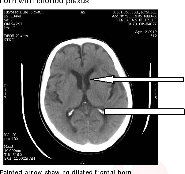
The Ventricular System and Cerebrospinal Fluid (CSF) Simple Tube Shape of Early CNS with fluid filled canal in middle In adults there is still a continuous canal running thru all levels of the adult CNS – it is just harder to follow. Book Fig. 1.19 Side & Frontal Views of Ventricles Remember – these represent fluid filled cavities in brain of 8 to 10 axial image slices of the brain were obtained without any overlap. 2.3. Method of Measuring the Brain Ventricle The lateral ventricles on CT are seen as two band-shaped structures with normal attenuation values. The third ventricle is amid-line structure which is shown between the thalami and the fourth ventricle is seen as an oval
The paired lateral ventricles (ventriculi laterales I and II) are two of these four chambers and are connected to the unpaired third and fourth ventricle through the foramen interventriculare. The following article gives an overview on the topography of the ventricles, their structure, as well as the production and re-absorption of liquor. Neurology: Ventricular system of the Brain. STUDY. PLAY. Human Brains-Brain is suspended in CSF and partially floats in the skull cavity -There are 4 cavities in the brain called ventricles. All 4 ventricles are interconnected through narrow-pipe like openings. CSF-is produced predominately in the brain ventricles -functions as a buffer to absorb stress transferred through the rigid skull
of 8 to 10 axial image slices of the brain were obtained without any overlap. 2.3. Method of Measuring the Brain Ventricle The lateral ventricles on CT are seen as two band-shaped structures with normal attenuation values. The third ventricle is amid-line structure which is shown between the thalami and the fourth ventricle is seen as an oval The most severe form of slit ventricle syndrome occurs in children. The absence of cerebrospinal fluid (CSF) within the ventricles combined with a growing brain leads to situation in which "the brain is too big for the skull." The intracranial pressure (brain pressure) can be very high.
Dandy W. E. Benign encapsulated tumors in the lateral ventricles of the brain. Diagnosis and treatment. Baltimore: Williams & Wilkins Co. 1934 viii 189 pp. Dandy W. E. Benign encapsulated tumors in the lateral ventricles of the brain. Diagnosis and treatment. Neurology: Ventricular system of the Brain. STUDY. PLAY. Human Brains-Brain is suspended in CSF and partially floats in the skull cavity -There are 4 cavities in the brain called ventricles. All 4 ventricles are interconnected through narrow-pipe like openings. CSF-is produced predominately in the brain ventricles -functions as a buffer to absorb stress transferred through the rigid skull
Hetzel W. The posterior horn and collateral trigone of the lateral ventricle of the monkey brain (Macaca speziosa). A scanning electron microscopic study. Cell Tissue Res. 1978 Jan 9; 186 (1):161–170. Hirunagi K, Yasuda M. Scanning electron microscopic analysis of the linings of the fourth ventricle in the domestic fowl. 28/03/2014 · This video is about the ventricles of the brain. For more videos visit seewhyanatomy.com or follow us on twitter @seewhyanatomy.
The ventricles are hollow spaces in the human brain.They contain cerebrospinal fluid (CSF). There are four ventricles in a human brain. This is the ventricular system: it continues as the central canal of the spinal cord.The ventricles are interconnected: this allows the flow of cerebrospinal fluid. N7 Ventricles of Brain 1. left lateral ventricle 2. right lateral ventricle 3. anterior horn 4. inferior horn 5. posterior horn 6. median portion of lateral ventricle 7. communication between lateral ventricle and third ventricle 8. third ventricle 9. interthalamic adhaesion 10. portion of third ventricle reaching into pineal gland
The most severe form of slit ventricle syndrome occurs in children. The absence of cerebrospinal fluid (CSF) within the ventricles combined with a growing brain leads to situation in which "the brain is too big for the skull." The intracranial pressure (brain pressure) can be very high. 22/02/2015 · Ventricles of the brain: want to learn more about it? Our engaging videos, interactive quizzes, in-depth articles and HD atlas are here to get you top results faster. Sign up for your free Kenhub account today and join over 1,195,034 successful anatomy students.
The present investigation examined the ependymal linings of the cerebral ventricles of the dog following a single intracisternal injection of the viable … The ventricular system is a series of connecting hollow spaces called ventricles in the brain that are filled with cerebrospinal fluid. The ventricular system consists of two lateral ventricles, the third ventricle, and the fourth ventricle. The cerebral ventricles are connected by small pores called foramina, as well as by larger channels.The interventricular foramina or foramina of …
Anatomy of meninges, ventricles, cerebrospinal fluid 1. MENINGES, VENTRICLES, CEREBROS PINAL FLUID AND BOOLD SUPPLY OF THE BRAIN Dr. Israa M. Sulaiman Dr. Mohammed Faez Department of Anatomy IMS/MSU 2. OBJECTIVES • Illustrate and describe the Meninges’s three membranes. • Describe the structure of the meninges, its blood supply and nerve Start studying Ventricular System of the Brain. Learn vocabulary, terms, and more with flashcards, games, and other study tools.
22/06/2013 · This brief animation illustrates the location of the ventricles within the brain and the associated structures around it. The structures were sculpted digitally using ZBrush and were combined with 29/05/2015 · Ventricles of the Brain-+ Dailymotion. For You Explore. Do you want to remove all your recent searches? All recent searches will be deleted. Cancel Remove. Log in. Watch fullscreen. Ventricles of the Brain. Windowbluff

Anatomy of meninges, ventricles, cerebrospinal fluid 1. MENINGES, VENTRICLES, CEREBROS PINAL FLUID AND BOOLD SUPPLY OF THE BRAIN Dr. Israa M. Sulaiman Dr. Mohammed Faez Department of Anatomy IMS/MSU 2. OBJECTIVES • Illustrate and describe the Meninges’s three membranes. • Describe the structure of the meninges, its blood supply and nerve The Ventricular System of the Brain - Free download as Powerpoint Presentation (.ppt / .pptx), PDF File (.pdf), Text File (.txt) or view presentation slides online. cns